Datasets:
Tasks:
Image Segmentation
Modalities:
Image
Formats:
parquet
Sub-tasks:
instance-segmentation
Languages:
English
Size:
1K - 10K
ArXiv:
License:
| dataset_info: | |
| features: | |
| - name: image | |
| dtype: | |
| image: | |
| mode: RGB | |
| - name: instances | |
| sequence: | |
| image: | |
| mode: '1' | |
| - name: categories | |
| sequence: | |
| class_label: | |
| names: | |
| '0': Neoplastic | |
| '1': Inflammatory | |
| '2': Connective | |
| '3': Dead | |
| '4': Epithelial | |
| - name: tissue | |
| dtype: | |
| class_label: | |
| names: | |
| '0': Adrenal Gland | |
| '1': Bile Duct | |
| '2': Bladder | |
| '3': Breast | |
| '4': Cervix | |
| '5': Colon | |
| '6': Esophagus | |
| '7': Head & Neck | |
| '8': Kidney | |
| '9': Liver | |
| '10': Lung | |
| '11': Ovarian | |
| '12': Pancreatic | |
| '13': Prostate | |
| '14': Skin | |
| '15': Stomach | |
| '16': Testis | |
| '17': Thyroid | |
| '18': Uterus | |
| splits: | |
| - name: fold1 | |
| num_bytes: 283673837.64 | |
| num_examples: 2656 | |
| - name: fold2 | |
| num_bytes: 267595457.439 | |
| num_examples: 2523 | |
| - name: fold3 | |
| num_bytes: 293079722.82 | |
| num_examples: 2722 | |
| download_size: 1665092597 | |
| dataset_size: 844349017.8989999 | |
| configs: | |
| - config_name: default | |
| data_files: | |
| - split: fold1 | |
| path: data/fold1-* | |
| - split: fold2 | |
| path: data/fold2-* | |
| - split: fold3 | |
| path: data/fold3-* | |
| license: cc-by-nc-sa-4.0 | |
| task_categories: | |
| - image-segmentation | |
| task_ids: | |
| - instance-segmentation | |
| language: | |
| - en | |
| tags: | |
| - medical | |
| - cell nuclei | |
| - H&E | |
| pretty_name: PanNuke | |
| size_categories: | |
| - 1K<n<10K | |
| paperswithcode_id: pannuke | |
| # PanNuke | |
| [](https://warwick.ac.uk/fac/cross_fac/tia/data/pannuke) | |
| ## Dataset Description | |
| - **Homepage:** [PanNuke Dataset for Nuclei Instance Segmentation and Classification](https://warwick.ac.uk/fac/cross_fac/tia/data/pannuke) | |
| - **Leaderboard:** [Panoptic Segmentation](https://paperswithcode.com/sota/panoptic-segmentation-on-pannuke) | |
| ## Description | |
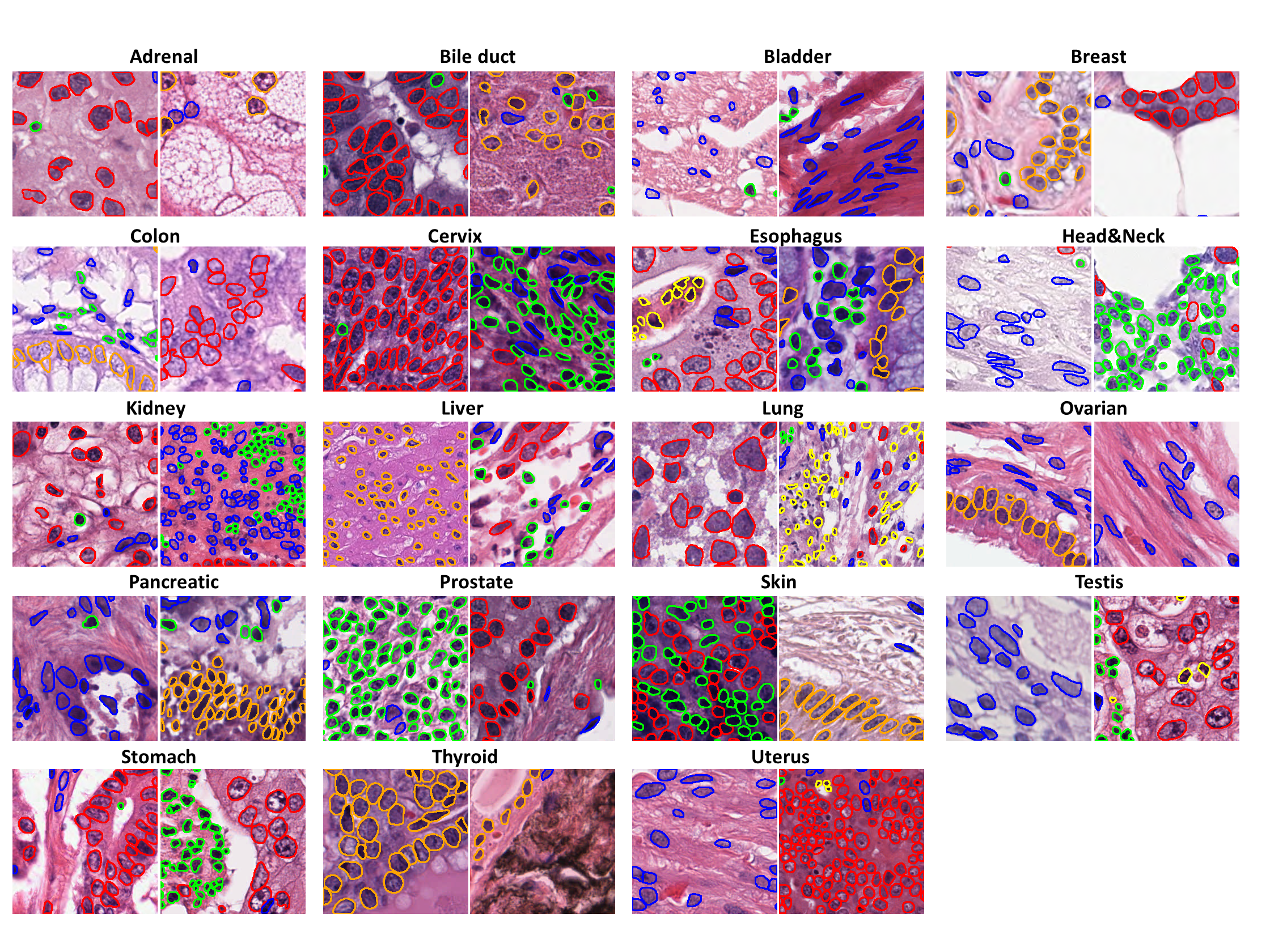
| PanNuke is a semi-automatically generated dataset for nuclei instance segmentation and classification, providing comprehensive nuclei annotations across 19 tissue types and 5 distinct cell categories. The dataset includes a total of **189,744 labeled nuclei**, each accompanied by an instance segmentation mask, and contains **7,901 images**, each sized **256×256 pixels**. The images were captured at **x40 magnification** with a resolution of **0.25 µm/pixel**. The dataset is highly imbalanced, with the **"Dead" nuclei category** being particularly underrepresented. | |
| Please note that the dataset was created by extracting patches from whole-slide images (WSIs). As a result, some nuclei located at the edges of patches may be cropped, with fewer than 10 visible pixels in certain cases. | |
| ## Dataset Structure | |
| The dataset is organized into three folds: `fold1`, `fold2`, and `fold3`, consistent with the original dataset structure. Each fold contains data in a tabular format with the following four columns: | |
| - **`image`**: The RGB tile of the sample. | |
| - **`instances`**: A list of nuclei instances. Each instance represents exactly one nucleus and is in binary format (`1` - nucleus, `0` - background) | |
| - **`categories`**: An integer class label for each nucleus, corresponding to one of the following categories: | |
| 0. Neoplastic | |
| 1. Inflammatory | |
| 2. Connective | |
| 3. Dead | |
| 4. Epithelial | |
| - **`tissue`**: The integer tissue type from which the sample originates, belonging to one of these categories: | |
| 0. Adrenal Gland | |
| 1. Bile Duct | |
| 2. Bladder | |
| 3. Breast | |
| 4. Cervix | |
| 5. Colon | |
| 6. Esophagus | |
| 7. Head & Neck | |
| 8. Kidney | |
| 9. Liver | |
| 10. Lung | |
| 11. Ovarian | |
| 12. Pancreatic | |
| 13. Prostate | |
| 14. Skin | |
| 15. Stomach | |
| 16. Testis | |
| 17. Thyroid | |
| 18. Uterus | |
| ## Citation | |
| ```bibtex | |
| @inproceedings{gamper2019pannuke, | |
| title={PanNuke: an open pan-cancer histology dataset for nuclei instance segmentation and classification}, | |
| author={Gamper, Jevgenij and Koohbanani, Navid Alemi and Benes, Ksenija and Khuram, Ali and Rajpoot, Nasir}, | |
| booktitle={European Congress on Digital Pathology}, | |
| pages={11--19}, | |
| year={2019}, | |
| organization={Springer} | |
| } | |
| ``` | |
| ```bibtex | |
| @article{gamper2020pannuke, | |
| title={PanNuke Dataset Extension, Insights and Baselines}, | |
| author={Gamper, Jevgenij and Koohbanani, Navid Alemi and Graham, Simon and Jahanifar, Mostafa and Khurram, Syed Ali and Azam, Ayesha and Hewitt, Katherine and Rajpoot, Nasir}, | |
| journal={arXiv preprint arXiv:2003.10778}, | |
| year={2020} | |
| } | |
| ``` |